STROKE

Patients Stroke

There are two subtypes of hemorrhagic stroke: intracranial hemorrhage (ICH) and subarachnoid hemorrhage (SAH). During a hemorrhage, due to the bleeding of blood vessels, normal supply of blood to brain tissues in parts of the brain are cut off. When there is bleeding directly into the brain tissue, often forming a clot, the stroke is defined as intracranial hemorrhage; When bleeding fills the cerebrospinal fluid spaces around the brain, the stroke is defined as subarachnoid hemorrhage. Both conditions are very serious, resulting in high mortality and disability rates.

What causes the disease?

Smoking, high blood pressure (hypertension), hardening of the arteries (arteriosclerosis), alcohol use, and underlying diseases which can increase the risk of developing brain aneurysms. Some people may be genetically prone to developing aneurysms which is why your physician will be interested in your family history.

Stroke Patients stroke1 1

Can hemorrhagic strokes be prevented?

While a hemorrhagic stroke cannot always be prevented, you can lower your chances of having a hemorrhagic stroke by:

  • Getting treatment for high blood pressure – This is very important, because untreated high blood pressure is a common cause of hemorrhagic strokes. Treatment can involve lifestyle changes, diet changes, and/or medication.
  • Not smoking.
  • Not using illegal drugs.

If an abnormal blood vessel was the cause of the stroke, surgery can sometimes be performed to prevent it from bleeding again.

Stroke Patients stroke2 2

Follow-up and recovery

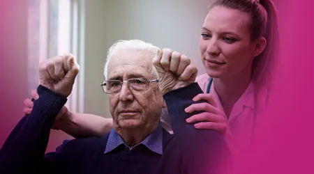
Some people recover from a stroke without any long-term impact to their quality of life or with only minor impact. However, many have seriously reduced functionality after a stroke. For example, they might be unable to speak or feed themselves, or they might be unable to move parts of their body. Specialists, such as physical therapists and speech therapists, can help.
Stroke Patients stroke3 3

Diagnosis

  1. Head CT and MRI scan: The importance of CT scanning in the early stage of onset is to exclude cerebral hemorrhage, but no abnormal findings will be found at early stage of ischemic stroke. Low-density changes can be detected 24–28 hours after onset of disease. MRI can identify disease 4 hours after onset.
  2. Cerebrovascular examination: Digital subtraction angiography (DSA), CT or MR angiography can show the location and nature of the stroke and help doctors to evaluate the disease before performing treatment.
  3. Transcranial Doppler (TCD): The degree of blockage of the cerebral vessels can be determined by the velocity and direction of blood flow.
Stroke Patients stroke4 4

What causes the disease?

There are two types of obstruction that results in ischemic stroke:

  • Cerebral Thrombosis: The formation of a blood clot in a cerebral vein in the brain, which is responsible for draining blood from the brain.
  • Cerebral Embolism: The formation of a blood clot elsewhere in the body, which makes its way into the brain’s blood vessels. Usually this blood clot comes from the heart or the body’s large arteries such as the upper chest or neck. Irregular heart beat (Atrial Fibrillation) and diseased heart valves can also increase the risk of experiencing an ischemic stroke.
Stroke Patients stroke5 1

Symptoms

  • Face: Does the person's face look uneven or droop to one side?
  • Arm: Does the person have weakness or numbness in one or both arms? Does one arm drift down if the person tries to hold both arms out?
  • Speech: Is the person having trouble speaking? Does his or her speech sound strange?
  • Time: If you see any of these signs, call emergency services immediately. You need to act FAST and get the person to a hospital. The sooner treatment begins, the better the chances of recovery.
Stroke Patients stroke6 2

Risks

  • High levels of cholesterol or triglycerides.
  • High blood pressure.
  • Diabetes.
  • Smoking.
  • Unhealthy eating habits.
  • Overweight or obesity.
  • Physical inactivity.
  • Family history of stroke.
  • Heart disease.
Stroke Patients stroke7 3

Diagnosis

  1. Head CT and MRI scan: The importance of CT scanning in the early stage of onset is to exclude cerebral hemorrhage, but no abnormal findings will be found at early stage of ischemic stroke. Low-density changes can be detected 24–28 hours after onset of disease. MRI can identify disease 4 hours after onset.
  2. Cerebrovascular examination: Digital subtraction angiography (DSA), CT or MR angiography can show the location and nature of the stroke and help doctors to evaluate the disease before performing treatment.
  3. Transcranial Doppler (TCD): The degree of blockage of the cerebral vessels can be determined by the velocity and direction of blood flow.
Stroke Patients stroke8 4